Advanced Childbirth Simulator
View DetailsAdvanced Childbirth Simulator
Part No:158
Features:
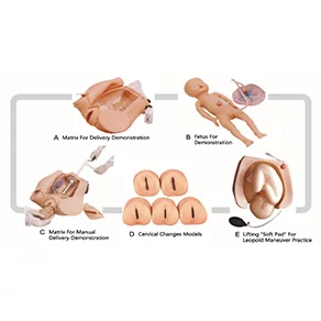
1. Demonstrate the fetus, umbilical cord and placenta of vacuum-assisted delivery, flexible fetal joint, and multiple normal and abnormal positions of fetal delivery can be showed;
2. Equipped with manual delivery mechanical parts; by manual , the whole delivery process, including engagement, descending, flexion, internal rotation, extension, restoration, external rotation and fetal delivery, can be showed;
3. Can practise and master the comprehensive skills of normal labor, abnormal labor (dystocia), midwifery and perineum protection;
4. Equipped with soft elevated cushion for Leopold Maneuver practice;
5. Equipped with models for cervical and vaginal examination practice before and at birth;
Component: B ( Fetus for Demonstration ) + C ( Matrix for Manual Delivery Demonstration ) +
D ( Cervical Changes Models ) + E ( Lifting "Soft Pad" for Leopold Maneuver Practice )
Leopold Maneuvers, Anal and Vaginal Examination Simulator
View DetailsLeopold Maneuvers, Anal and Vaginal Examination Simulator
Part no :160
Features:
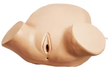
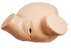
1. The simulation is made from good material with vivid appearance, soft and elastic texture and realistic feeling.
2. Allow to aerate the abdomen to adjust upheaval, and have Leopold's maneuvers training and assessment.
3. Allow to have vaginal and anal examination to ascertain the fetal position.
4. Pelvic measurement.
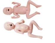
Thirty-two Weeks Premature Infant Simulator
View DetailsThirty-two Weeks Premature Infant Simulator
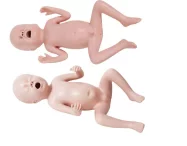
Part No: 168
Features:
The product has physiological features of premature infant. Weight is less than 2500g; body length is shorter than 47cm. Retained testicle of male infant; female infant’s labium majus can not cover labium minus; visible of physiologic icterus. Provide bregmatic palpation practice, premature infant bathing and care in the incubator. Allow premature infant suction and nasal feeding practice. The model features realistic feeling, soft texture, good flexibility and durability, can be practiced for many times.
GD/FT331B thirty-two Weeks Premature Infant Model
32 weeks of pregnancy (7.5 months)
Body weight 1700g (1300-2100g)
Body length 39cm (38-42cm)
Application range
1. The model is designed as the teaching tool of O & G examination demonstration and exercise operation training. It can be used for clinical education demonstration and students practice training for medical schools, nursing college and medical college.
2. For hospital clinical Gynecology and Obstetrics staff education practice operation;
3. For the clinical medical popularity training in the basic health unit;
Maintenance and warranty
1. After training session, wipe the model clean and encase, then put it in the dry and cool place so as to extend the using life.
2. with spare parts for replacement.
3. We guarantee a period of two years free warranty basing on the copy of purchasing invoice and warranty card; lifetime maintenance, and cost should be liable for users.
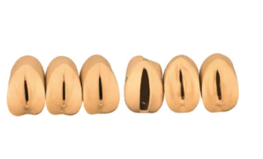
Prenatal Cervical Change Model
View DetailsPrenatal Cervical Change Model
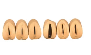
Part No:162
For who use:
1. The models are suitable for the training in medical college, nursing school and clinic training institute.
2. They can be used for the training of nurse, intern student, etc.
3. Grass-root medical institute.
Function and Guideline:
1. The group of models has six parts. Vaginal speculum can be used in the observation of the variation of prenatal cervix and birth canal. The inspection of the variation of pre-delivery can also be performed in the models. The inspector should wear sterile gloves before operation, meanwhile, inspector should use French chalk to lubricate the fingers in the glove as well as lubricate the vaginal speculum and the surface of the models.
2. There are six phases in the course of prenatal cervix variation:
Phase 1: The orifice of cervix has no expanding, and the cervical tunnels haven't disappearedat all. The positional relation between the head of fetus and the square of ischial spine is -5.
Phase 2: The orifice of cervix has expanded 2cm bigger, and the cervical tunnels disappear 50%. The positional relation between the head of fetus and the square of ischial spine is -4.
Phase 3: The orifice of cervix has expanded 4cm bigger, and the cervical tunnels disappear absolutely. The positional relation between the head of fetus and the square of ischial spine is -3
Phase 4: The orifice of cervix has expanded 5cm bigger, and the cervical tunnels disappear absolutely. The positional relation between the head of fetus and the square of ischial spine is 0.
Phase 5: The orifice of cervix has expanded 7cm bigger, and the cervical tunnels disappear absolutely. The positional relation between the head of fetus and the square of ischial spine is +2.
Phase 6: The orifice of cervix has expanded 10cm bigger, and the cervical tunnels disappear absolutely. The positional relation between the head of fetus and the square of ischial spine is +5.
Maintenance and Storage:
1. In case of damage, Lubricant should be applied when people practise in the models.
2. Impregnant and corrosive can cause damage, so get rid of the materials during practise. Don't use ballpoint pen to make mark when practise. To clean the surface of the model, people can use soap or warm water, then use napkin or soft cloth tomop up the remain liquid.
3. there are spare parts for replacement.
4. For long-time storage, clear and dry the models. And then store them in a ventilative and dry place.
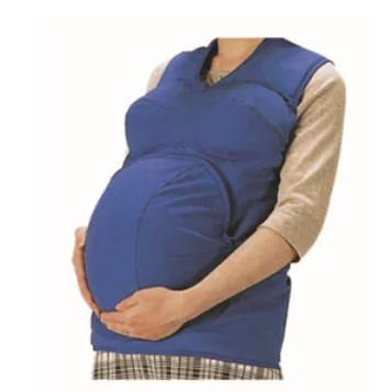
Advanced Wearable Gravida Simulator
View DetailsAdvanced Wearable Gravida Simulator
Part No:162
Features:
1. The model is applicable to male adult, unmarried female, student and premarital education and learning;
2. 13kgs abdominal weight can realistically simulate 40weeks of pregnancy; can also simulate different weeks of pregnancy via adjusting the liquid in the uterine cavity;
3. Wearable design, can carry out four maneuvers of Leopold, breast nursing and other prenatal diagnosis;
4. Wearing it and experience chest distress, lumbago, urinary frequency and other physiological state because of hysterauxesis during
gestation period; can also experience pregnant women’ all kinds of inconvenience;
5. Inject warm water into uterus to simulate amniotic fluid, can also measure uterus height and abdominal circumference;
6. Can be adjusted to be close to the best condition of human body, and act bloom sampling (8-10 weeks of pregnancy) and amniotomy (16-20 weeks of pregnancy).
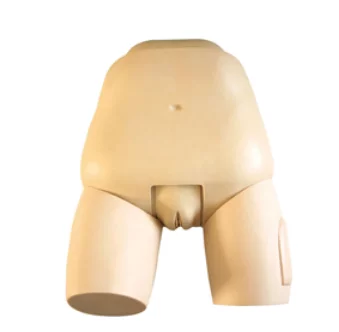
Advanced Fundus of Uterus Examination & Evaluation Simulator
View DetailsAdvanced Fundus of Uterus Examination & Evaluation Simulator
Part No:162
Features:
1. With normal physiological changes of the postpartum women, provide assessment of postpartum fundus of uterus and massage skills training;
2. Hip joint is movable and can be placed correct body position.
3. Realistic anatomical landmark of united bones of toes;
4. Interchangeable Uterus:
Normal uterus
Postpartum uterus of the first hour
Postpartum uterus of the second day
Postpartum uterus of the second week
Callous uterus with good contraction
Soft uterus without good contraction
5. Expansion of vaginal orifice
6. Easy to separate the labium minus and expose the vagina,
7. Lear energy of cunnus
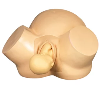
Vacuum Delivery Simulator
View DetailsVacuum Delivery Simulator
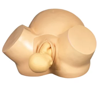
Part No:162
Features:
1. Can demonstrate all the standard delivery procedures.
2. Consistent with the real pelvic verity size and with main anatomical marks.
3. Fetus model: the skin is soft and fontanel is visible, vacuum extraction can be exercised.
4. Flexible fetus joints fetal position through transformation demonstrate a variety of normal and abnormal position childbirth.
Size:59cm*39cm*41.5cm
G.W.:6.5kg
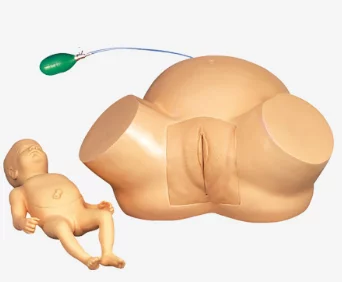
Diffcult Labor Simulator
View DetailsDiffcult Labor Simulator
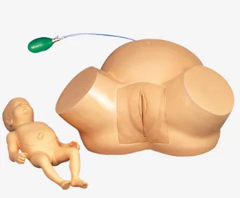
Part No:162
Features:
1. Can demonstrate strate all the situation of abnormal birth.
2. Inflatable demonstrate the narrow of the pelvis.
3. Simulate the abnormal fetal position, can process presentation dystocia.
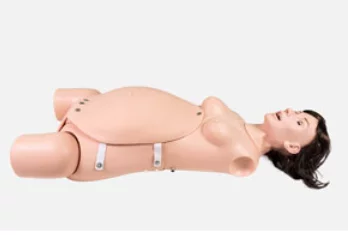
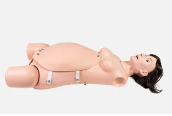
Advanced Ostetrics Surgery Training Manikin
View DetailsAdvanced Ostetrics Surgery Training Manikin
Part No:162
Features:
1. This product is a half thigh model in the lithotomy position, which can be used for obstetric surgery simulation training.
2. It can carry out various teaching and skill training such as episiotomy, artificial peeling and membrane rupture, cesarean section, uterine gauze packing, and uterine rupture repair.
3. It is equipped with an arterial blood circulation system, and simulated blood is ejected when the blood vessels are damaged by surgery, which makes the training, teaching and assessment of surgical skills more realistic.
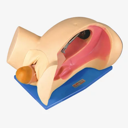
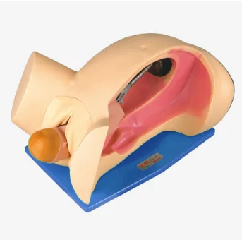
Delivery Mechanism Demonstration Simulator
View DetailsDelivery Mechanism Demonstration Simulator
Part No:164
Features:
1. Vertical section model, designed in accordance with the human physiological anatomy structure.
2. Accurate anatomic marks, obvious outline of uterus and bony pelvis.
3. Delivery mechanical parts demostrate the whole delivery process, inclduing engagement, descending, flexion, internal rotation, extension, resoration, external rotation and shoulder delivery.
4. Simulational birth canal skin, simulate the real delivery situation.
5. The fetal skin is soft and fontanel is visible.
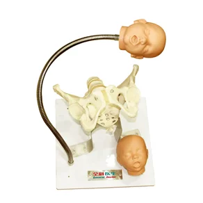
Pelvis with Fetal Heads Model
View DetailsPelvis with Fetal Heads Model
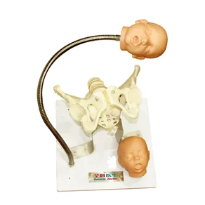
Part No:164
Features:
1. Consist of a female pelvis and two fetal heads;
2. Two fetal heads: one is full-term, the other is premature;
3. Each cranial sutures and anterior and posterior fontanels are palpable on fetal skull;.
4. Fetal heads are fixed on flexible shaft through the pelvis;
5. Can observe the position relation of the fetus and pelvis during delivery;
6. Intuitionally demonstrate the birth process of engagement, descending, flexion, internal rotation, extension, restoration, external rotation and fetal delivery;
7. Can demonstrate forceps and vaccum assisted deliveries.
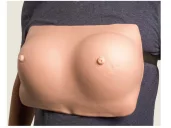
Breast Examination Simulator
View DetailsBreast Examination Simulator
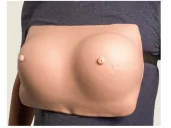
Part No: 166
Features:
Palpation of malignant tumor, benign tumor, lymphaden and lobulus accrementition.
Size:37cm*18.5cm*29.5cm
G.W.:4kg
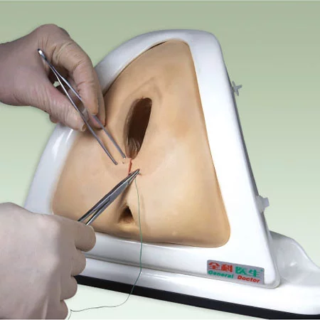
Episiotomy Traning Simulator
View DetailsEpisiotomy Traning Simulator
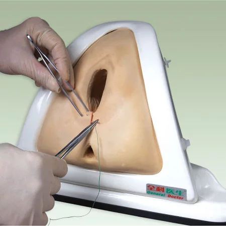
Part No:164
Features:
1. This model is designed for practice of perineum cutting and suturing.
2. The main structure of the simulator is a perineum, which is made of simulative foam, and the inner muscle is soft PVC. The simulator is mounted on a plastic framework.
3. There are two handles in the back and bottom of the simulator to adjust tension of the perineum.
4. Right posterior incision, left posterior incision or median incision can be selected to exercise perineum cutting and suturing.
Size:36cm*34cm*26cm
G.W.:2kg
Advanced Inspection and Palpation of Breast Simulator
View DetailsAdvanced Inspection and Palpation of Breast Simulator
Part No :166
Features:
1. This model can be used to exercise mammary tu mors palpation skill.
2. In order to facilitate training process, we implant benign tumors and malignant tumors into the mammary gland.
3. Basing on various position, size, texture, the students can dentify the tumor is benign or malignant.
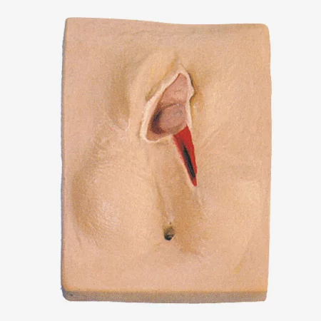
Vulva Suturing Training Simulator
View DetailsVulva Suturing Training Simulator
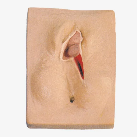
Part No:164
Features:
Right posterior incision, left posterior incision or median incision can be selected to exercise perineum cutting and suturing.
Size:19.5cm*21cm*25cm
G.W.:1.2kg
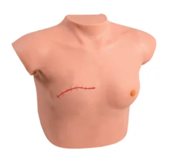
Model for Breast Examination and Postoperative Care of Mammectomy
View DetailsModel for Breast Examination and Postoperative Care of Mammectomy
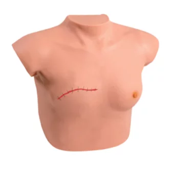
Part No: 166
Features:
The model could be laid flat on table for teaching demonstration. It also could be hung in front of the chest. Conduct breast lumps palpation by sitting or dorsal position. Lumps of benign and malignant tumor are put into each side of mammary tissue to make palpation more convenient. It is available to identify comparatively and make a definite diagnosis through position, size, texture, and features like moving or fixed on the basis of palpation. The model is very useful for differential diagnosis of benign lump and malignant tumor of breast. Moreover, it is available to perform postoperative care of mammectomy of one side.
Vaginal Culdocentesis Simulator
View DetailsVaginal Culdocentesis Simulator
Part No:164
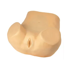
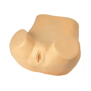
The model consists of cortex, fixed abdominal visceral organs, uterus, uterine-rectal concave haematodocha, vagina, rectal water sac and the stand. The cortex is used as the cover of the whole model; the devices used in belly are to fix functional visceral organs on their own actual locations. And four parts (uterine-rectal concave water sac, rectal water sac, uterus and vagina) are inlayed into the device. The stand can hold the whole model and keep its breech up, which makes operation simple. The uterine-rectal concave haematodocha and rectal water sac are of cystic structure where to connect two drainage tubes to have liquid infused into.
Features:
1. The simulation model is made from advanced materials with correct anatomical positions, soft and elastic skin, true feeling and real pathological tissue;
2. Penetrate into median fornix posterior, which is1 cminferior to the joint of cervix and vagina mucosa, parallel to cervical canal with a No.7 puncture needle, and light red liquid extraction means successful centesis;
3. If puncture isn’t operated according to the conventions, e.g. if the operator pierces into the rectum, there will be yellow liquid extraction, which shows puncture failure;
4. If operator doesn’t insert the needle according to the convention but pierces bilaterally without destination, it will hurt surrounding organs, which shows puncture failure.
Gynecological Examination Simulator
View DetailsGynecological Examination Simulator
Part No:164
Features:
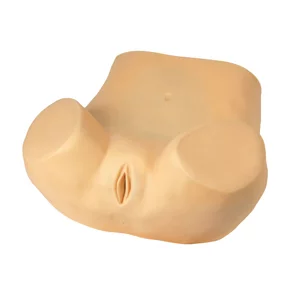
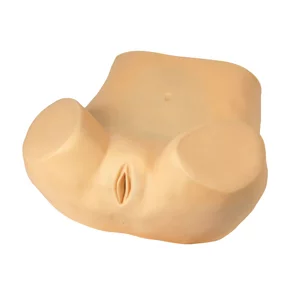
1. Palpation of normal and pregnant uterus
2. Bi-manual pelvic and tri-manual pelvic examination
3. Examine the normal cervix via vaginoscope and coplposcope
4. Place and remove the IUD
5. Pregnant uterus and postpartum uterus examination
6. Observe the uterus, ovaries, fallopian, round ligament and other anatomy structure of pelvic.
7. Anteposition and retroposition of uterus can be adjusted
8. Pelvic measurement.
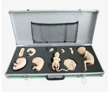
Advanced Embryo Development Process Simulator
View DetailsAdvanced Embryo Development Process Simulator
Part No: 166
Features:
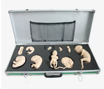
1. Be placed in the beautiful rigid box, divided into 9 different process of embryo development
2. Videos vividly describe the embryonic development, the magical process of embryonic growth and reveal the mysteries of life.
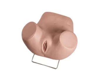
Advanced Gynecological Examination Simulator
View DetailsAdvanced Gynecological Examination Simulator
Part No :166
Features:
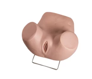
1. It is including all the functions of GD/F30S.
2. Hysteroscopy checking
3. Laparoscopic visualization checking, can observe the Uterus, accessories, round ligament, uterine and other structures
4. It can place the female condoms, contraceptive sponge, cervical cap and other contraceptives.
5. Cervix, uterus can be replaced
The components:
The normal and abnormal uterus and accessories model can be bimanual and triple diagnosis checking.
1) Normal uterus and accessories (the front uterus wall is transparent, so the IDU can be placed and took out.)
2) Normal uterus and accessories
3) Uterus associated with significant flexion, forward.
4) Uterus associated with significant back flexion, back forward.
5) Uterus associated with right ovarian cysts tubal.
6) Uterus associated with right tubal cyst.
7) Uterus associated with right hydrosalpinx
8) Uterine fibroids
9) Uterine inflammation associated with the right accessories uterine malformations.
Normal and abnormal cervix model (it is used for checking the parts by gynecatoptron)
1) Normal cervix
2) Chronic cervicitis
3) Acute cervicitis
4) Cervical cyst inflammation Bohnert
5) Cervical polyps
6) Cervical adenoma
Normal and abnormal cervix model (it is used for checking the parts by Hysteroscopy)
1) Normal uterus
2) Endometrial polyps
3) Endometrial hyperplasia
4) Uterine fibroids
5) Early uterine body cancer
6) Advanced uterine body cancer
7) End uterine cancer
8) Pregnant uterus model
9) 6-8 weeks of pregnancy the uterus
10) 10-12weeks of pregnancy the uterus
11) 20 weeks of pregnancy the uterus
12) Simulation of post partum 48 hours uterus
13) The process of placing and taking out the IDU
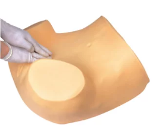
Gynecological Examination Simulator
View DetailsGynecological Examination Simulator
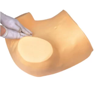
Part No :166
Features:
1. Palpation of normal and pregnant uterus
2. Bi-manual pelvic and tri-manual pelvic examination
3. Vaginal examination
4. Visual recognition of normal and abnormal cervices
5. Contraceptive instrument insertion and removal
6. Observation of uterus, ovary, fallopian tubes, and ligament teres
7. Normal and abnormal uterus and attachment model
8. Place and remove IUD by using the guide fork of contraceptive ring
9. Gravid uterus(Five-mouth fetus )
10. Extra-uterine pregnancy(ampullary pregnancy)
11. Salpingemphraxis
Size:68.5cm*29cm*55.5cm
G.W.:12.5kg
